Female Chest Muscle Anatomy Diagram - Women Strength Training Exercise Muscle Map Transformation Body Muscle Anatomy Strength Training Workouts - This article looks at female body parts and their functions, and it provides an interactive diagram.
Female Chest Muscle Anatomy Diagram - Women Strength Training Exercise Muscle Map Transformation Body Muscle Anatomy Strength Training Workouts - This article looks at female body parts and their functions, and it provides an interactive diagram.. Its origin is the sternum and median ventral raphe, and its insertion is at the humerus. Quadratus lumborum quadratus lumborum is actually a muscle of the posterior wall, but it is often described as part of the ventral trunk musculature. It is smaller than the pectoralis minor muscle. May 31, 2021 reading time: And the ovaries, which produce the anatomically female egg cells.
This muscle originates from the iliac crest and iliolumbar ligament. On the belly you will see the umbilical cord which connected the fetal pig to its mother's placenta. It is smaller than the pectoralis minor muscle. Molly smith dipcnm, mbant • reviewer: Its action is to draw the arm towards the chest.
Chest Muscles Anatomy Woman Anatomy Drawing Diagram from img-new.cgtrader.com However, most of its anterior border is covered by the pectoralis major. It contracts and flattens when you inhale. Quadratus lumborum quadratus lumborum is actually a muscle of the posterior wall, but it is often described as part of the ventral trunk musculature. The pectoralis minor muscle is larger than the pectoralis major. Its action is to draw the arm towards the chest. Nov 05, 2019 · female anatomy includes the external genitals, or the vulva, and the internal reproductive organs. May 31, 2021 reading time: May 31, 2021 · muscle anatomy reference charts author:
Molly smith dipcnm, mbant • reviewer:
Brain anatomy provide the labels for the diagram on the left below and provide descriptions of the functions of each structure on the blank lines. Its origin is the sternum and median ventral raphe, and its insertion is at the humerus. This article looks at female body parts and their functions, and it provides an interactive diagram. Dimitrios mytilinaios md, phd last reviewed: Jul 30, 2018 · the diaphragm is a thin skeletal muscle that sits at the base of the chest and separates the abdomen from the chest. It is smaller than the pectoralis minor muscle. Mar 18, 2015 · the major muscle in the chest is the pectoralis major. The pectoralis minor muscle is larger than the pectoralis major. The axial region makes up the main axis of the human body and includes the head, neck, chest, and. Molly smith dipcnm, mbant • reviewer: On the belly you will see the umbilical cord which connected the fetal pig to its mother's placenta. However, most of its anterior border is covered by the pectoralis major. In the diagram to the left, provide the labels for the structures involved in the reflex act when a person steps on a tack and jerks their leg away.
And the ovaries, which produce the anatomically female egg cells. Regional terms describe anatomy by dividing the parts of the body into different regions that contain structures that are involved in similar functions. In the diagram to the left, provide the labels for the structures involved in the reflex act when a person steps on a tack and jerks their leg away. However, most of its anterior border is covered by the pectoralis major. Dimitrios mytilinaios md, phd last reviewed:
Female Body Front Surface Anatomy Human Body Shapes Anterior View Parts Of Human Body General Anatomy The Anatomical Canstock from comps.canstockphoto.com This muscle originates from the iliac crest and iliolumbar ligament. Brain anatomy provide the labels for the diagram on the left below and provide descriptions of the functions of each structure on the blank lines. Dimitrios mytilinaios md, phd last reviewed: On the belly you will see the umbilical cord which connected the fetal pig to its mother's placenta. Mar 18, 2015 · the major muscle in the chest is the pectoralis major. Regional terms describe anatomy by dividing the parts of the body into different regions that contain structures that are involved in similar functions. Its origin is the sternum and median ventral raphe, and its insertion is at the humerus. Two primary terms are used to describe the main regions of the body:
May 31, 2021 reading time:
Brain anatomy provide the labels for the diagram on the left below and provide descriptions of the functions of each structure on the blank lines. Regional terms describe anatomy by dividing the parts of the body into different regions that contain structures that are involved in similar functions. This article looks at female body parts and their functions, and it provides an interactive diagram. On the belly you will see the umbilical cord which connected the fetal pig to its mother's placenta. In the diagram to the left, provide the labels for the structures involved in the reflex act when a person steps on a tack and jerks their leg away. Quadratus lumborum quadratus lumborum is actually a muscle of the posterior wall, but it is often described as part of the ventral trunk musculature. However, most of its anterior border is covered by the pectoralis major. It is smaller than the pectoralis minor muscle. May 31, 2021 · muscle anatomy reference charts author: Dimitrios mytilinaios md, phd last reviewed: The female reproductive system contains two main parts: Its origin is the sternum and median ventral raphe, and its insertion is at the humerus. Mar 18, 2015 · the major muscle in the chest is the pectoralis major.
This muscle originates from the iliac crest and iliolumbar ligament. Its origin is the sternum and median ventral raphe, and its insertion is at the humerus. Regional terms describe anatomy by dividing the parts of the body into different regions that contain structures that are involved in similar functions. The axial region makes up the main axis of the human body and includes the head, neck, chest, and. In the diagram to the left, provide the labels for the structures involved in the reflex act when a person steps on a tack and jerks their leg away.
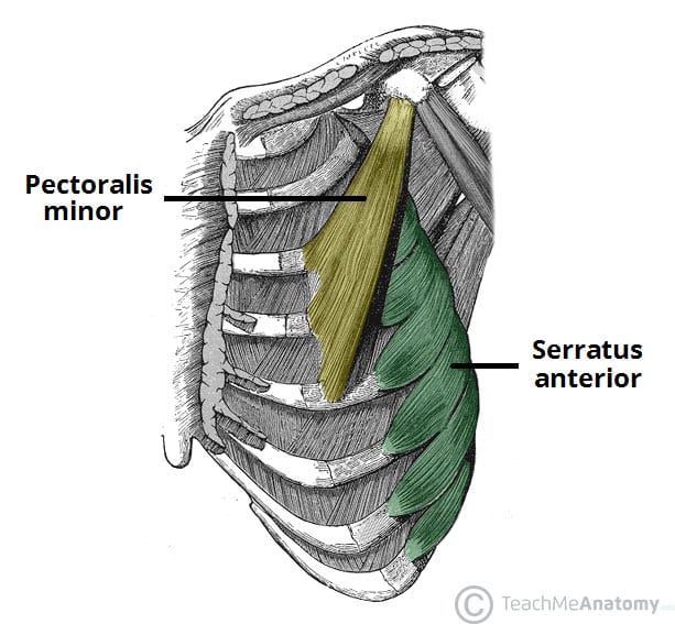
Muscles Of The Pectoral Region Major Minor Teachmeanatomy from teachmeanatomy.info In the diagram to the left, provide the labels for the structures involved in the reflex act when a person steps on a tack and jerks their leg away. The female reproductive system contains two main parts: The axial region makes up the main axis of the human body and includes the head, neck, chest, and. Brain anatomy provide the labels for the diagram on the left below and provide descriptions of the functions of each structure on the blank lines. And the ovaries, which produce the anatomically female egg cells. This article looks at female body parts and their functions, and it provides an interactive diagram. It is smaller than the pectoralis minor muscle. Mar 18, 2015 · the major muscle in the chest is the pectoralis major.
The axial region makes up the main axis of the human body and includes the head, neck, chest, and.
In the diagram to the left, provide the labels for the structures involved in the reflex act when a person steps on a tack and jerks their leg away. The female reproductive system contains two main parts: Two primary terms are used to describe the main regions of the body: Brain anatomy provide the labels for the diagram on the left below and provide descriptions of the functions of each structure on the blank lines. May 31, 2021 reading time: Its origin is the sternum and median ventral raphe, and its insertion is at the humerus. The pectoralis minor muscle is larger than the pectoralis major. The axial region makes up the main axis of the human body and includes the head, neck, chest, and. Mar 18, 2015 · the major muscle in the chest is the pectoralis major. Dimitrios mytilinaios md, phd last reviewed: It contracts and flattens when you inhale. Regional terms describe anatomy by dividing the parts of the body into different regions that contain structures that are involved in similar functions. It is smaller than the pectoralis minor muscle.
On the belly you will see the umbilical cord which connected the fetal pig to its mother's placenta chest muscle anatomy diagram. Mar 18, 2015 · the major muscle in the chest is the pectoralis major.


0 Komentar